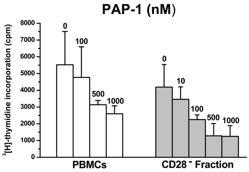
Figure 3.

PAP-1 suppresses the proliferation of PBMCs and TEM cells from RM. PBMCs isolated from healthy uninfected RM (n = 9) were seeded at 100,000 cells/well and were stimulated with 400 ng/ml plate-bound anti-CD3 in the absence or presence of increasing concentrations of PAP-1 (nM). To test the effect of PAP-1 on TEM cells, PBMCs were depleted of CD28+ cells using anti-CD28-coated magnetic microbeads and the CD28− fraction was seeded at 100,000 cells/well. Cultures were incubated at 37°C, 7% CO2 for 3 days. Cells were pulsed with 1 uCi/well [3H]-thymidine 16 hrs prior to harvest and thymidine uptake was measured by standard scintillation counting. The mean cpm ± S.D was determined; shown is data representative of three independent assays.
