
Fig 5.
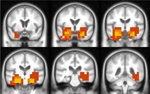
Anatomic patterns with maximum discriminative power between AD and controls are overlaid on the corresponding custom T1 template. Color scale used to indicate the occurrence of the voxel in multiple tissue maps. Yellow: voxel location used in all three tissues (GM, WM and CSF); orange: voxel location used in at least two tissues and red: voxel location used in one tissue only.