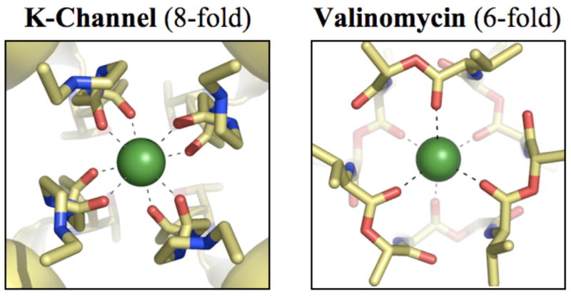
Figure 1.
Structural differences between K+ binding sites in strongly selective K-channels and valinomycin. (a) Partial view of the x-ray structure9 of a representative K-channel, KcsA, illustrating a K+ ion occupying the S2 site of the selectivity filter in a state of high coordination by 8 carbonyl oxygens. (b) X-ray structure36 of valinomycin illustrating a K+ ion coordinated by 6 carbonyl oxygens. K+ ions are drawn as green spheres and all other atoms as sticks with oxygens in red, carbons in yellow and nitrogens in blue. Dashed lines connecting the ion and the oxygen atoms represent coordination with K+.

