
Fig. 2. Detail of subcellular localization of Epac-1 or Rap1 near phagocytic cup.
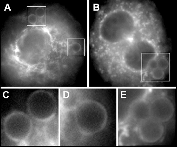
Following phagocytosis of opsonized beads for 60 min, AMs were fixed and stained for Epac-1 (A, C, D) or Rap1 (B, E). Images in C and D are details from A, and E is a detail of the indicated area in image B.