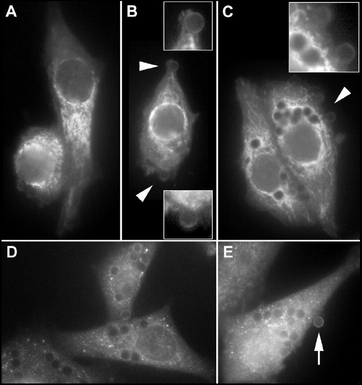
Fig. 3. Subcellular localization of Epac-1, Rap1 and GAPDH in the AM cell line NR8383.

Cells were incubated without beads (A) or with opsonized beads for 15 (B) or 60 (C-E) min. Insets in (B) and (C) are details of the regions indicated by arrowheads. Arrow in (E) indicates peripherally bound bead with positive staining for GAPDH.
