
Fig. 5. Localization of Epac-1 and Rap1 following phagocytosis of opsonized beads in AMs pretreated with PGE2.
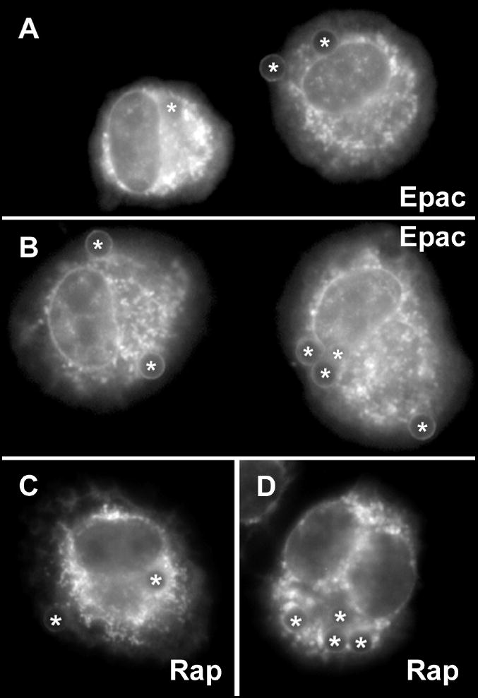
Cells were pretreated with 1 μM PGE2 for 15 min and then incubated with opsonized beads for 60 min. Two fields from independent experiments are presented for Epac-1 (A, B) and Rap1 (C, D)). Asterisks (*) indicate phagosomes containing beads.