
Figure 5. DNA Contacts.
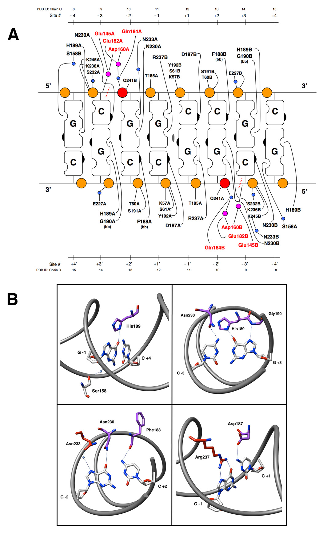
Panel A: Contacts make by the NotI restriction endonuclease to its 8 basepair DNA recognition sequence. Orange circles represent the phosphate groups of the DNA sugar-phosphate backbone. The scissile phosphats are colored red. Mounds and indents on the cartoon blocks represent the hydrogen bond donors and acceptors, respectively, of the individual nucleotide bases. The right hand side of the bases represents the major groove of the DNA and the left side represents the minor groove. The 2-fold rotation axis in the center of the cartoon designates the center of symmetry for the palindromic target DNA sequence. Small blue circles represent water molecules, and magenta circles are calcium ions. Residues from the active site are colored red. The site of DNA cleavage is designated by a dashed red line. The bases of the recognition sequence are numbered both outward from the center of symmetry (designated Site #) as well as by their PDB ID from the model coordinates. Panel B: Specific contacts made to each basepair of the NotI recognition sequence. Protein sidechain stick representations are colored based on their domain of origin: clamp domain (purple) or DNA recognition helix (red). Important atoms of bases and sidechains are colored by element. Hydrogen bonding interactions are represented by dashed lines. Waters are shown as blue spheres.