
Fig. 7.
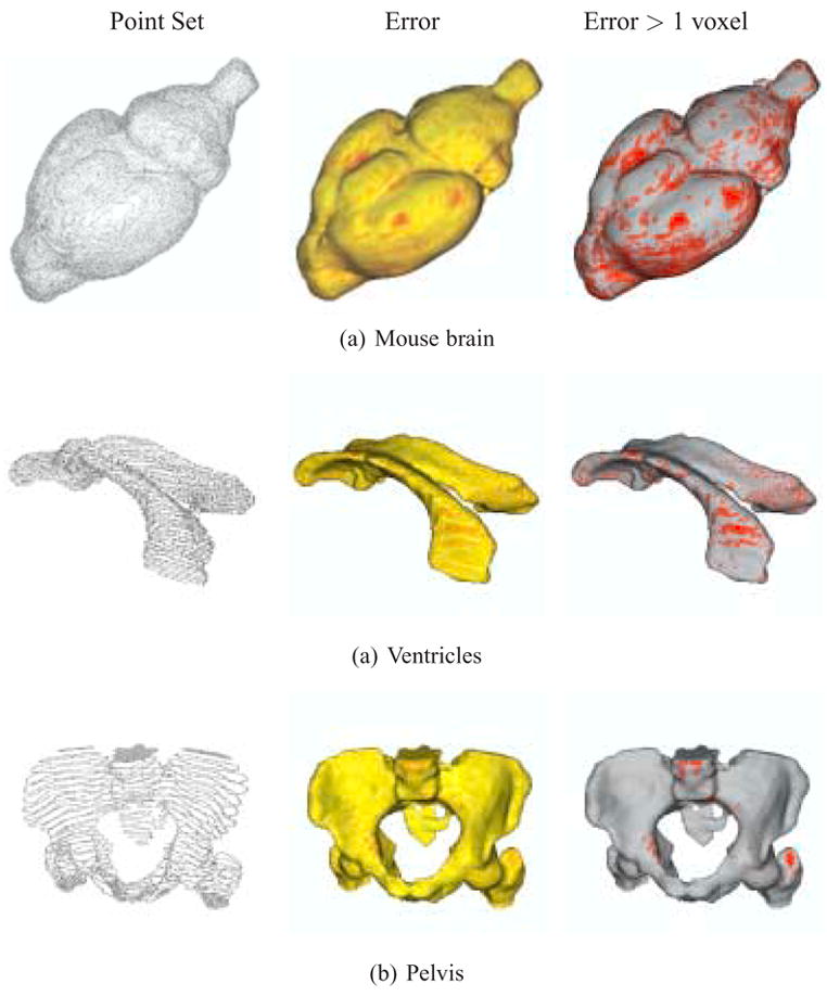
Reconstruction of anisotropic mouse brain, ventricles and pelvis data, with approximation errors. (left) Point set extracted from the input contours. (center) Reconstruction error from yellow (low) to red (high). (right) Regions with errors greater than 1 voxel marked in red.