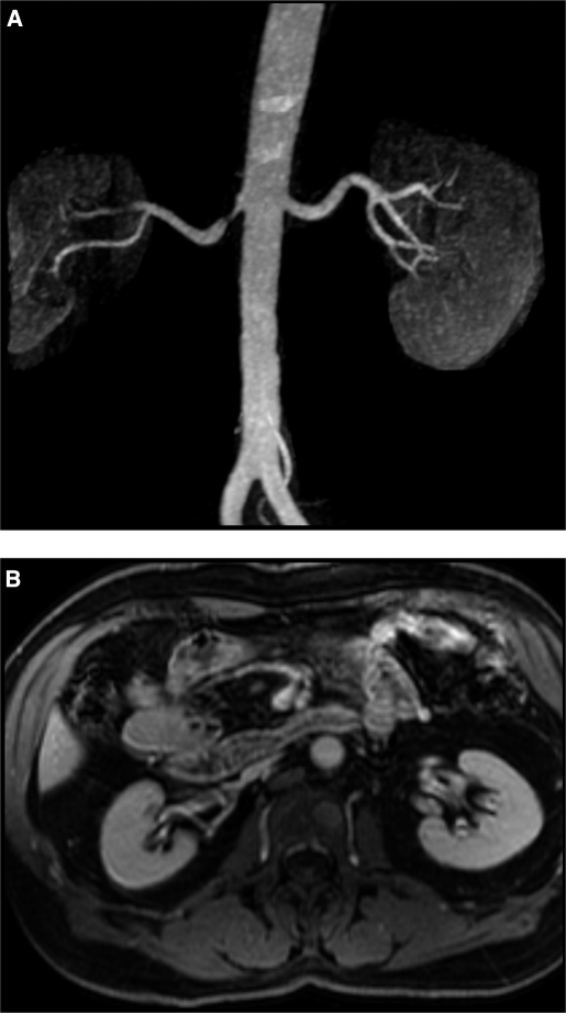
Figure 2.
Magnetic resonance (MR) angiogram (A) demonstrating focal stenosis of the proximal right renal artery. Despite the stenosis and poststenotic dilation, filtration and kidney volume (B) were preserved in this kidney. This is an example of a “normal” appearing kidney beyond a stenotic lesion (see text). The left kidney in this patient was considered a “normal” kidney.

