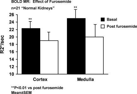
Figure 3.
BOLD MR measurements (R2*, s−1) in cortex and medullary segments in 21 “normal” appearing kidneys before and after intravenous furosemide. Relative reduction in cortical segments (11.2 ± 2%) was less than that observed in medullary regions (20 ± 2%) (P < 0.05). Enhanced reductions in R2* after furosemide in medullary segments is consistent with relatively greater accumulation of deoxyhemoglobin related to oxygen consumption due to chloride and sodium transport in the thick ascending limb of Henle (see text).

