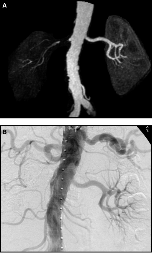
Figure 4.
MR angiogram (A) demonstrating near total occlusion to the right kidney with minimal filtration. Conventional intra-arterial contrast angiography (B) (one week later) confirmed total occlusion and nonfunction of this kidney. This is an example of “nonviable” kidney as summarized in Table 2.

