Figure 4.

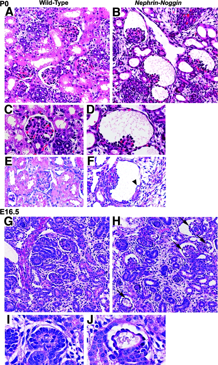
Histologic analysis of Nephrin-Noggin mice at P0 and E16.5. (A through F) Kidneys from wild-type (A, C, and E) and Nephrin-Noggin (B, D, and F) mice at P0 (periodic acid-Schiff staining). Nephrin-Noggin mice developed collapse of glomerular capillary tufts and dilation of Bowman's capsule (B, D, and F). Tubule population was decreased in Nephrin-Noggin mice (B) compared with wild-type mice (A). Some cystic glomeruli in Nephrin-Noggin mice had rudimentary tubules (arrowhead in F). (G though J) Embryonic kidneys at E16.5 from wild-type (G and I) and Nephrin-Noggin (H and J) mice (periodic acid-Schiff staining). Overall structure of the kidney was comparable between wild-type (G) and Nephrin-Noggin (H) mice. At this stage, neither cystic glomeruli nor loss of tubules was observed in Nephrin-Noggin mice (H). Microaneurysms were formed in glomerular capillary tufts of Nephrin-Noggin mice (H and J, arrows), which were absent in wild-type mice (G and I). Magnifications: ×200 in A, B, G, and H; ×400 in C, D, E, F, I, and J.
