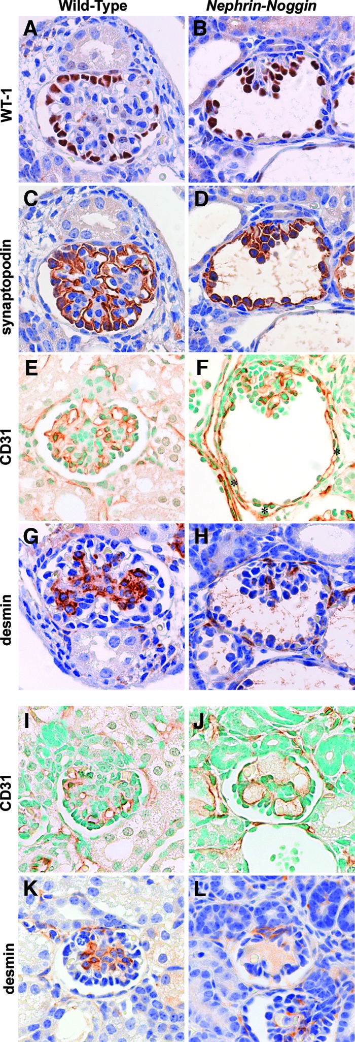
Figure 5.

Immunohistologic analysis of Nephrin-Noggin mice. Immunostaining for WT-1 (A and B), synaptopodin (C and D), CD31 (E, F, I, and J), or desmin (G, H, K, and L) in glomeruli of wild-type (A, C, E G, I, and K) or Nephrin-Noggin (B, D, F, H, J, and L) mice at birth. (A through H) Glomeruli were from deep cortical area of kidneys of wild-type (A, C, E, and G) or Nephrin-Noggin (B, D, F, and H) mice. Glomeruli of Nephrin-Noggin mice showed cystic change with collapse of capillary tuft. In wild-type mice, intense WT-1 staining was confined to glomerular visceral epithelial cells (A), whereas it was observed in both visceral and parietal epithelial cells in Nephrin-Noggin mice (B). Similarly, synaptopodin was stained only in visceral epithelial cells in wild-type mice (C), whereas it was stained in both visceral and parietal epithelial cells in Nephrin-Noggin mice (D). Total number of WT-1–or synaptopodin-positive cells and their intensity in each cell were similar between the two strains of mice. (E) CD31 staining depicts glomerular and peritubular capillaries in wild-type mice. In Nephrin-Noggin mice, collapsed glomeruli contain CD31-positive cells. In addition, intense CD31 staining was ectopically observed just outside Bowman's capsule (F, asterisks). (G) Desmin staining depicts mesangial cells in wild-type mice. (H) The number of desmin-positive cells was markedly decreased in collapsed capillary tufts. (I through L) Glomeruli from the superficial cortex of wild-type (I and K) and Nephrin-Noggin (J and L) mice. Glomeruli of Nephrin-Noggin mice showed microaneurysms (J and H). They contained CD31-positive endothelial cells but almost no desmin-positive mesangial cells (L). Magnification, ×400.
