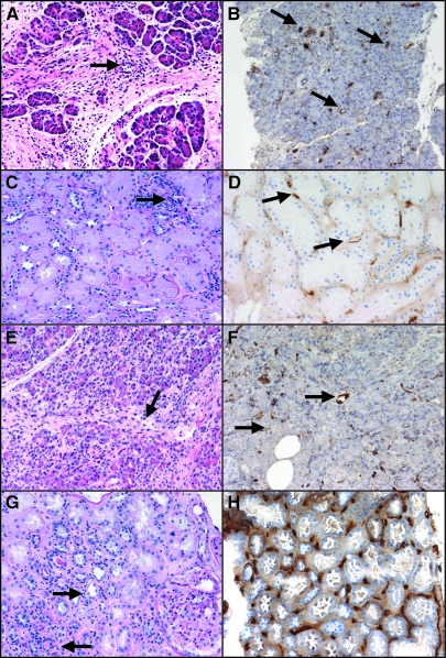
Figure 2.
Histopathology of the allografts in a patient with kidney and pancreas AMR. (A) Light micrograph of the transplant pancreas (postoperative day [POD] 155) shows moderate septal mononuclear inflammatory infiltrate with eosinophils and venous endothelialitis (arrow), grade II pancreas acute rejection (hematoxylin and eosin). (B) C4d immunolabeling of the same POD 155 pancreas biopsy reveals interacinar capillaries diffusely positive for C4d (arrows). (C) Periodic acid-Schiff stain section of the transplant kidney (POD 376) shows mild acute tubular injury, mild interstitial mononuclear inflammation (i1), and no tubulitis (t0). There is focal mild capillary margination of leukocytes (PTC score 1, arrows23). (D) C4d focally positive in the same POD 376 kidney biopsy (arrows). (E) Light micrograph of the transplant pancreas (POD 467) shows moderate septal and interacinar (arrows) mononuclear infiltrates with focal endothelialitis (arrow), grade III pancreas acute rejection (hematoxylin and eosin). (F) Interacinar capillaries diffusely positive for C4d in the same POD 467 pancreas biopsy). (G) Light micrograph of the transplant kidney 4 d later (POD 471) shows again mild interstitial inflammation with leukocyte margination (arrows) and no tubulitis. (H) Same POD 471 biopsy shows peritubular capillaries diffusely positive for C4d. Magnifications: ×200 in A and C through G; ×100 in B.

