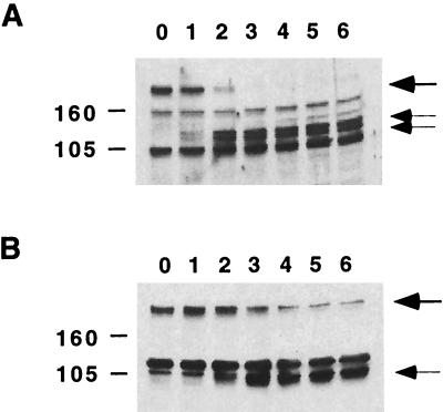
Figure 1.
Cleavage of eIF4GI and eIF4GII during poliovirus 3NC202 infection. Cell lysates (20 μg) collected at different times (hours; indicated at the top of the figure) after infection were separated by SDS/PAGE and were immunoblotted with antibodies directed against eIF4GI (A) or eIF4GII (B) (1, 27). Thick arrows indicate the migration of full-length eIF4G proteins, and thin arrows indicate the migration of the cleaved eIF4G products. Migration of known molecular weight markers is shown on the left.

