
Figure 3.
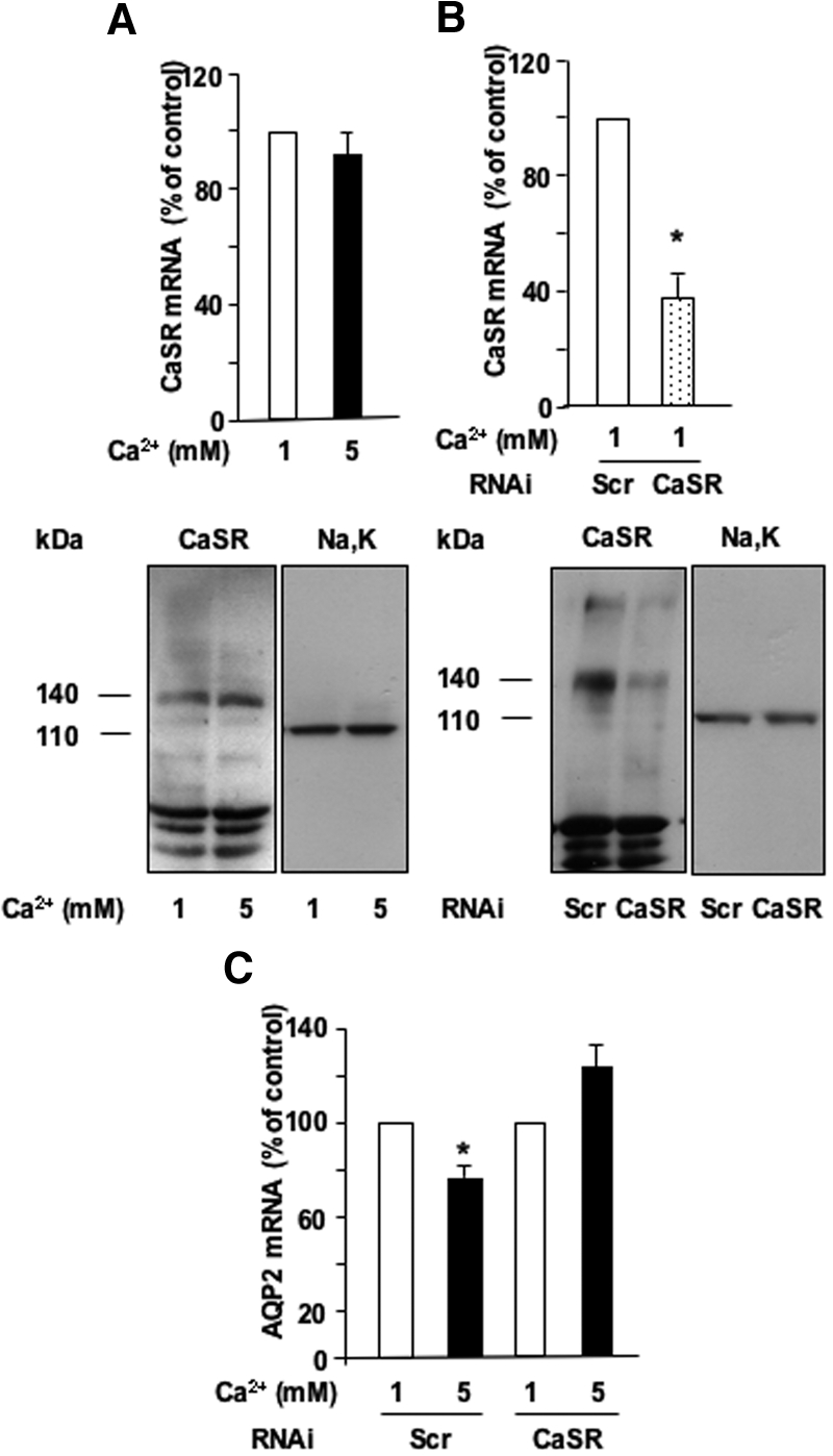
Role of the CaSR in the calcium-induced attenuation of AVP-dependent AQP2 expression. (A) Confluent mpkCCDcl4 cells grown on filters in culture medium containing baseline (1 mM) calcium were preincubated at 37°C with 10−9 M AVP for 24 h and then incubated in the continuous presence of AVP for another 24 h with 1 or 5 mM apical calcium. RNA or protein was then extracted and real-time PCR or Western blotting was performed as described in the Concise Methods section using primers or antibodies specific for CaSR, respectively. (B and C) Two days after transfection with either CaSR scramble RNAi (Scr) or CaSR RNAi, confluent mpkCCDcl4 cells grown on filters in culture medium containing baseline (1 mM) calcium were preincubated at 37°C with 10−9 M AVP for 24 h and then incubated in the continuous presence of AVP for another 14 h with 1 or 5 mM apical calcium. RNA or protein was then extracted and real-time PCR or Western blotting was performed as described in the Concise Methods sections using primers or antibodies specific for CaSR, respectively (B), or primers specific for AQP2 (C). Na-K-ATPase α-subunit was used as a loading control for Western blotting, and representative immunoblots from two independent experiments are shown. CaSR and Na,K-ATPase α-subunit were detected as 140- and 110-kD bands, respectively. PCR results are expressed as a percentage of control values determined after incubation in the presence of 1 mM calcium (100%). Bars are means ± SEM from six independent experiments. *P < 0.05.