
Figure 4.
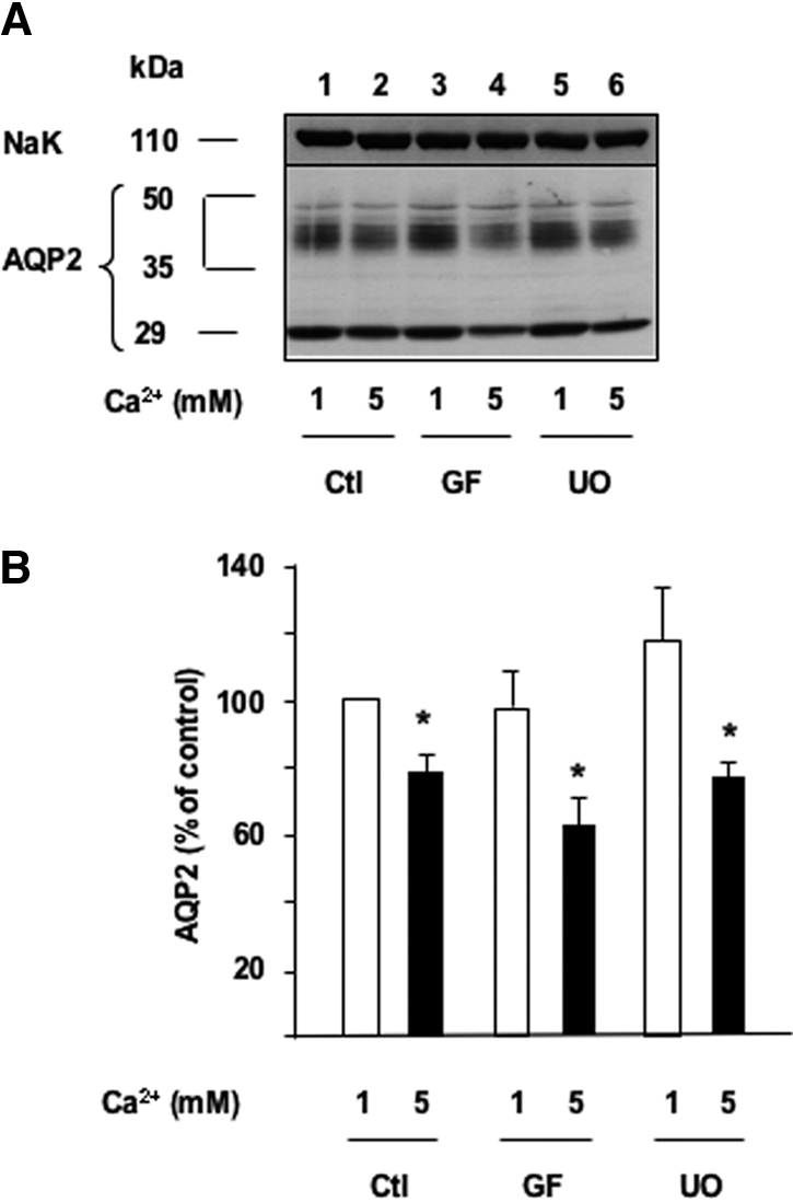
The effect of apical calcium on AVP-dependent AQP2 expression is independent of PKC and ERK activation. Confluent mpkCCDcl4 cells grown on filters in culture medium containing baseline (1 mM) calcium were preincubated for 24 h at 37°C with 10−9 M AVP. Cells were then incubated for another 24 h in the continuous presence of AVP and in the presence of 1 or 5 mM apical calcium and treated or not (Ctl) with either 10−6 M of the GF109203X (GF), a PKC inhibitor, or 10−6 M UO126 (UO), a MEK-1 inhibitor. Total protein extracts (40 μg) were separated by 10% SDS-PAGE and AQP2 and the Na-K-ATPase α-subunit, used as a loading control, were detected by Western blotting. (A) A representative immunoblot is shown. (B) Densitometric quantification of AQP2 protein expressed as a percentage of OD values measured in the presence of 1 mM calcium and without drugs (100%). Bars are means ± SE from four independent experiments. *P < 0.05.