
Figure 6.
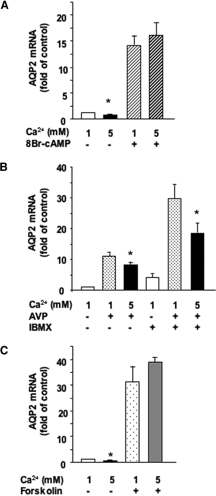
Role of phosphodiesterases and AC in the calcium-induced attenuation of AVP-dependent AQP2 expression. (A) Confluent mpkCCDcl4 cells grown on filters were preincubated for 24 h at 37°C in the presence of 1 or 5 mM apical calcium before incubation for another 24 h with or without 10−3 M 8Br-cAMP, a cell-permeable cAMP analog. (B) Confluent mpkCCDcl4 cells grown on filters were preincubated for 24 h at 37°C with 10−9 M AVP. Cells were then incubated for another 24 h in the continuous presence of AVP and in the presence of 1 or 5 mM apical calcium with or without 10−4 M IBMX, a phosphodiesterase inhibitor. (C) Confluent mpkCCDcl4 cells grown on filters were preincubated for 24 h at 37°C in the presence of 1 or 5 mM apical calcium before incubation for another 24 h with or without 5 × 10⁻⁶ M forskolin, a direct activator of AC. After RNA extraction, real-time PCR was performed as described in the Concise Methods section using primers against AQP2. Results are expressed as a percentage of control values determined after 24 h of incubation in the presence of 1 mM calcium and in the absence of drugs (100%). Bars are means ± SEM from four to eight independent experiments. *P < 0.05.