Figure 2.
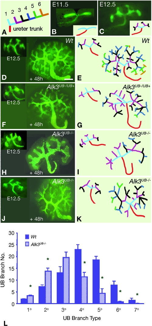
Primary ureteric bud branch pattern is disrupted in Alk3UB−/− kidneys. (A) Ureteric bud branches are classified according to their spatial orientation relative to the ureter trunk. (B and C) Green fluorescent protein (GFP) fluorescence in E11.5 and E12.5 Wt kidneys demonstrates the archetypal 1° ureteric bud branch pattern consisting of a ureter trunk (red) connected to two 1° ureteric bud branches (cyan). (D through K) GFP fluorescence of ureteric bud trees at E12.5 and Dolichos biflorus (DBA) fluorescence of these ureteric bud trees after 48 h of culture (+48h), together with corresponding color-coded stick figures. (D and E) The Wt (+48h) ureteric bud tree maintains the 1° branch pattern and exhibits 2° to 6° branch generations. (F and G) Some E12.5 Alk3UB−/UB+ kidneys exhibit a trifid branch pattern (inset in F) that is maintained after 48 h of culture. (H through K) Representative images of the trifid 1° branch pattern observed in the majority of Alk3UB−/− kidneys. (H and I) In some Alk3UB−/− (+48h) kidneys, severe reductions in 3° to 7° branch number and in total ureteric bud branch number are observed. Ureteric bud trunk and tip segments are also dilated, and terminal ureteric bud ampullae formed 4 to 6 new tips. (J and K) Some Alk3UB−/− (+48h) kidneys formed new 1° ureteric bud branches at the junction of the ureter trunk and initial ureteric branches. (L) Quantitation of ureteric bud branch number demonstrates that increased 1° to 2° ureteric bud branch number is associated with decreased 4° to 7° ureteric bud branch number in Alk3UB−/− (+48h) kidneys. Scale bar (D through K) = 100 μm.

