Figure 3.
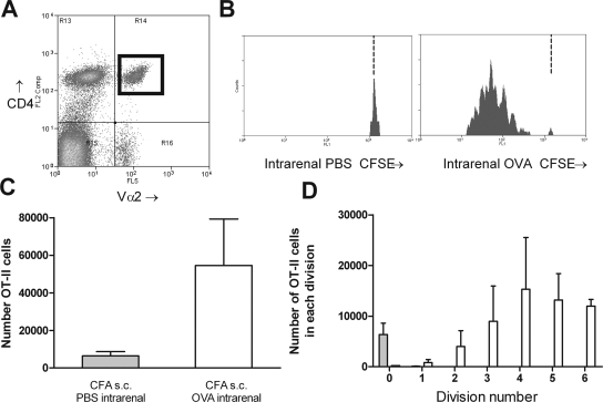
Intrarenal injection of OVA with subcutaneous CFA induces proliferation of naïve OT-II cells. A discrete population of CD4+Vα2+ cells was identifiable in recipients 72 h after OVA injection (A; CD4+Vα2+ cells marked by a black box in top right quadrant). This region allows identification of proliferating antigen-specific cells by CFSE fluorescence. The level of CFSE expression of these cells was assessed (B; illustrative single-parameter histograms). In mice injected with PBS intrarenally, CFSE+ cells were almost all expressing high levels of CFSE, indicating no proliferation. In OVA-injected mice, a series of peaks were identified indicating the serial halving of CFSE. The dotted vertical lines in B represent the degree of CFSE intensity of nonproliferating cells. CFSE− recipient cells (recipient CD4+Vα2+ cells, fluorescence intensity ≤11) were removed for clarity in Figures 3 through 6. After OVA injection, total numbers of OVA cells in the draining LN were increased (C), and the numbers in each division could be determined (D). □, Mice injected with OVA intrarenally, CFA subcutaneously; □, mice injected with PBS intrarenally, CFA subcutaneously.

