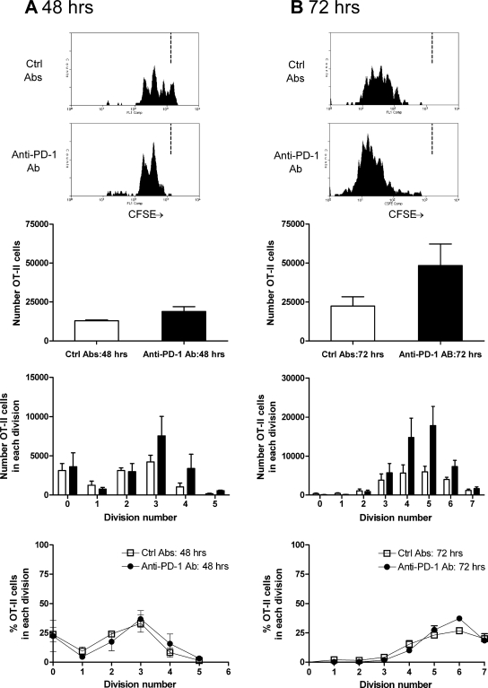
Figure 7.
The effect of PD-1 blockade on antigen-specific OT-II cell proliferation 48 h (A) and 72 h after intrarenal OVA injection (B). At 48 h, proliferation has commenced in mice injected with control Ab, and there is some effect of PD-1 at this early time point. After 72 h, the effect is more pronounced. Consistent with the lack of expression of PD-1 on naïve CD4+ cells, similar proportions of cells were in each division, suggesting that the time to first division is not affected by PD-1. Because these studies were performed by transferring CD45.2+ OT-II cells into congenic CD45.1 mice, the single-parameter histograms are from cells that are CD45.2+CD4+Va2+. The dotted vertical lines in the single-parameter histograms (A and B) represent the degree of CFSE intensity of nonproliferating cells. □, Mice injected with control Ab; ▪, mice injected with anti–PD-1 Ab.

