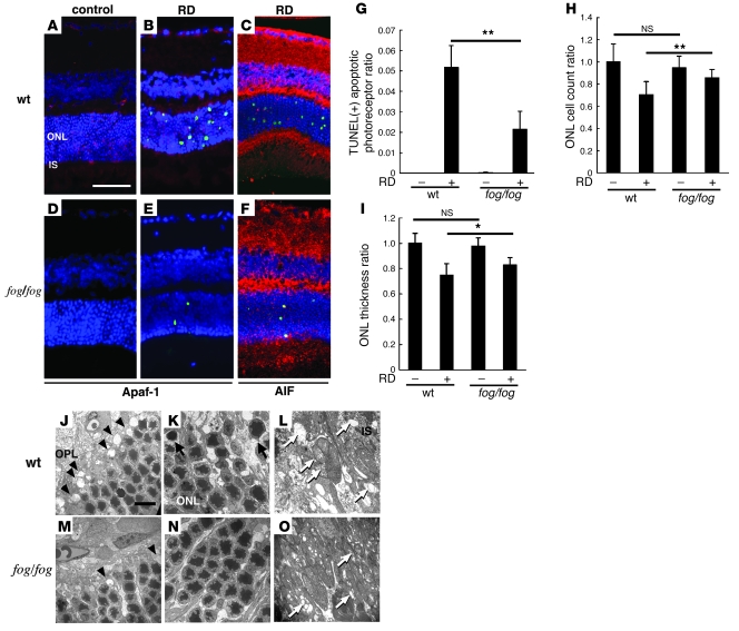
Figure 4. Apaf-1 deficiency protects from RD-induced photoreceptor apoptosis.
Apaf-1 expression was observed in WT mice (A and B; Apaf-1 in red, TUNEL in green, DAPI in blue) but deficient in Apaf-1 mutant fog/fog mice (D and E). TUNEL-positive apoptotic photoreceptor after RD decreased in fog/fog mice in contrast to WT mice (A–G). The remaining TUNEL-positive apoptotic photoreceptors showed AIF translocation into the nucleus in fog/fog mice (C and F; AIF in red, TUNEL in green, DAPI in blue). In fog/fog mice, ONL cell count ratio (H) and ONL thickness ratio (I) were also preserved, in contrast to those in WT mice. Ultrastructural studies showed relatively well-preserved structures in the outer plexiform layer (J and M, arrowheads; rod spherules and cone pedicles), the ONL (K and N, arrows; apoptotic photoreceptors), and the inner segment of photoreceptors (L and O, white arrows; ruptured mitochondria). n = 5 per group; *P < 0.05, **P < 0.01. Scale bars: 50 μm (A), 10 μm (J).

