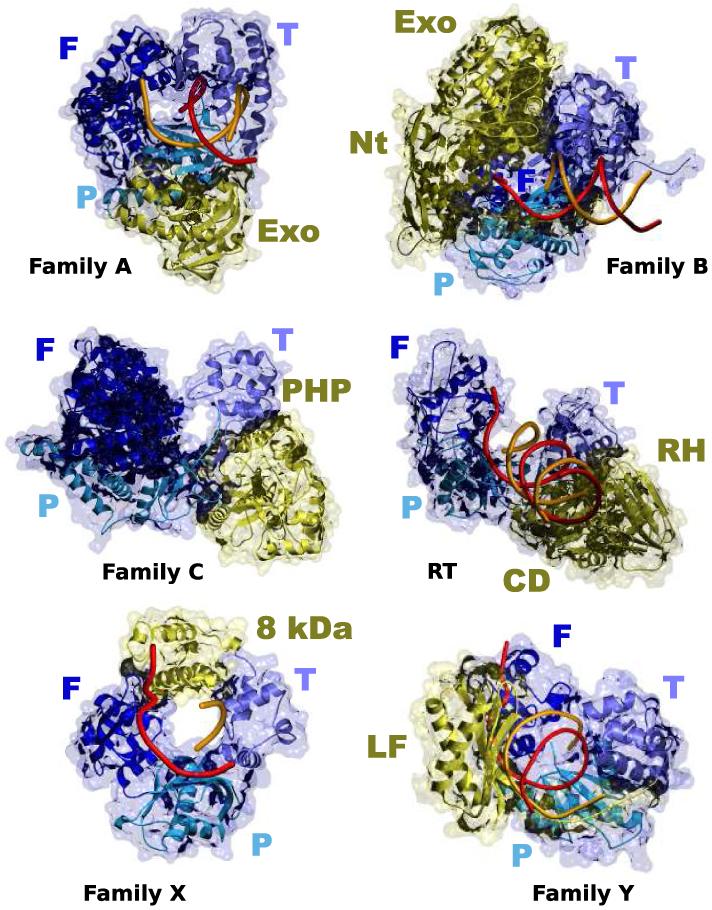
Figure 1. Structural similarity of DNA polymerases.

Crystal structures of a representative member of each of the five DNA polymerase families (A, T. aquaticus Pol I; B, RB69 Pol; C, E. coli Pol III; X, Pol λ and Y, S. solfataricus Dpo4) and retrotranscriptases (RT). All DNA polymerases contain a general fold that can be likened to a right hand, with fingers, palm and thumb subdomains, colored in different shades of blue. Additional subdomains, colored yellow, are specific for each family or individual enzyme. Exo, 3′-5′ exonuclease domain; Nt, N-terminal domain; PHP, Polymerase and Histidinol Phosphatase domain; 8 kDa, 8 kDa domain; LF, Little Fingers domain; RH, RNAse H domain; CD, Connecting domain.
