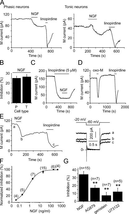
Figure 3.
NGF inhibited M/KCNQ currents from SCG neurons. (A) NGF (20 ng/ml) inhibited M/KCNQ currents from both phasic (left) and tonic (right) neurons. Time course of tail M/KCNQ currents at −60 mV was shown. Linopirdine (30 μM) was used to establish the baseline for the measurements. Perforated patch method was used in these experiments. (B) Summary data for NGF-induced inhibition of M/KCNQ currents shown in A. (C) NGF (20 ng/ml) inhibited M/KCNQ currents in presence of 5 μM linopirdine. (D) Oxo-M (10 μM) inhibited M/KCNQ currents. (E) NGF (20 ng/ml) also inhibited M/KCNQ currents recorded using conventional whole-cell method. The right panel shows the current traces taken at the times indicated in the left panel. (F) Concentration–response relationship for NGF-induced inhibition of M/KCNQ current. (G) Summary data for the effects of AG879 (50 μM, 5 min), genistein (100 μM, 5–10 min), and U73122 (3 μM, 3–5 min) on NGF-induced inhibition of M/KCNQ currents recorded using conventional whole-cell method. **, P < 0.01. Error bars represent SEM.

