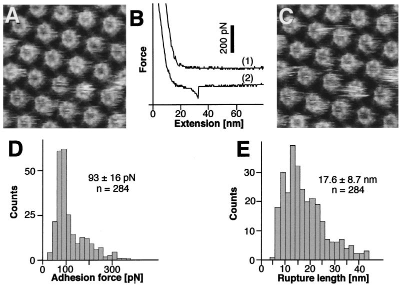
Figure 2.
Adhesion forces detected between the AFM stylus and the inner surface of the HPI layer. (A) Control AFM topograph of the inner surface of the HPI layer. The hexameric cores with their emanating arms have a central pore that can reversibly switch from a plugged to an unplugged conformation with a central plug (4, 18). The distance between the core centers is 18 nm. (B) On retraction of the stylus after indenting the protein with a loading force of 1 nN, 75% of the force-extension curves were similar to those recorded on the outer surface (trace 1; compare with Fig. 1B). The major fraction of the remaining force-extension curves (type I) showed an adhesion force of about 100 pN. Rupture occurred after extension of the molecular bridge by more than 10 nm (trace 2). (C) The same inner surfaces imaged after recording force curves; no structural changes occurred. The defect core on the right was used to align topographs C and A. (D) Histogram of the adhesion forces measured from 284 extension curves. The peak is at 93 ± 16 pN. (E) The corresponding distances between the stylus and the protein surface at the rupture point have a wide length distribution with an average of 17.6 ± 8.7 nm. Full gray-level range of the topographs corresponds to 3 nm.

