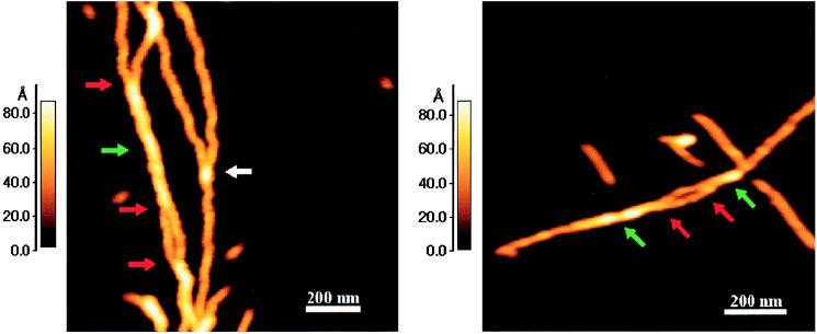
Figure 6.
Examples of branched fibrils indicating that they are composed of intertwined protofibrils. Both panels show fibrils (indicated by green arrows) that have a maximum height of 8.8 and a minimum height of 6.8 nm. Red arrows represent the regions in which two protofibrils (maximum height of 4.5 nm) were observed to branch out of the fibril. The regions in between the two red arrows in both the panels represent short portions of protofibrils that have come apart and are not intertwined. The white arrow represents a false branch point at which one protofibril simply lies on top of another and there is no intertwining.

