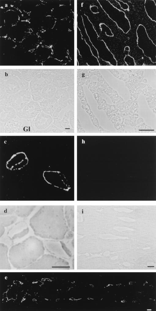
Figure 1.
Immunolocalization of PV-1 by immunofluorescence on semithin frozen section from different rat organs. Fluorescence and phase-contrast micrographs from: kidney (a and b), pancreas (c and d), intestine (e), adrenal glands (f and g), and heart (h and i), respectively. (a and b) The label is found specifically on the fenestrated peritubular capillaries. Please note the absence of signal from the glomerular capillaries (Gl) (where the endothelium is fenestrated but the fenestrae do not have diaphragms) or other renal structures. (c and d) An example of two labeled pancreatic acinary capillaries that shows the label on both sides of the cell. (e) Characteristic labeling of the fenestrated capillaries from an intestinal villus. (f and g) Labeled capillaries from the adrenal cortex. (h and i) Capillaries from the heart myocardium do not show any staining for PV-1. (Bars = 10 μm.)

