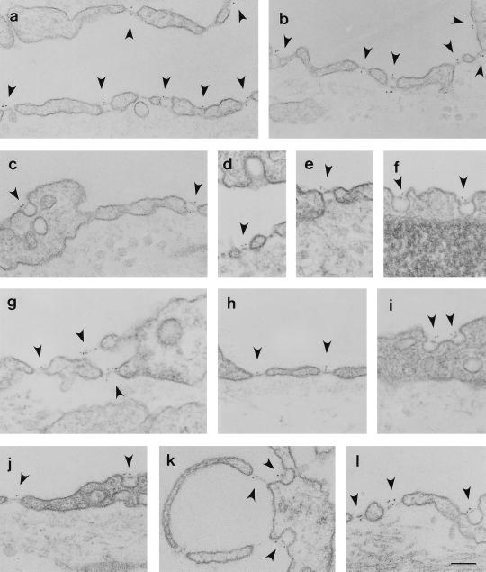
Figure 2.
Localization of PV-1 at EM level on the capillaries from kidney (a–c), intestine villi (d–f), adrenal cortex (g, k, and l), and pancreas (h–j). The respective tissues were labeled by using the anti-PV-1C polyclonal antibody followed by a 5-nm gold-conjugated reporter antibody as described in the text. Please note the labeling (arrowheads) of the stomatal diaphragms of transendothelial channels on both fronts of the endothelial cell. The fenestral diaphragms of the “sieve plates” or those of endothelial pockets (k) are labeled on both sides of the diaphragm. There is a remarkable lack of label on the proper (a–l), coated pits (d), or intercellular junctions (a). (Bar = 100 nm)

