Figure 2.
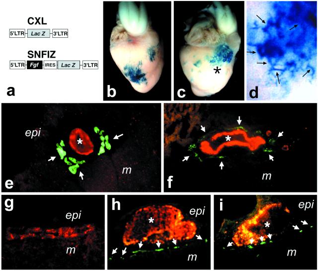
Ectopic induction of Purkinje fibers at FGF-induced sites of hypervascularization. (a) Proviral structures of replication-defective retroviruses expressing only β-gal (8, 14) or coexpressing FGF4 and β-gal (14, 17) used for infection of coronary precursors in vivo on E3 (16, 18). (b and c) Hearts infected with control β-gal virus (b) or FGF-producing virus (c). (d) FGF-induced vessels (some prominent branches are arrowed) adjacent to the location indicated by the asterisk in c. (e–i) Cryosections double-labeled with ALD58 (green) and anti-β-gal (red) or anti-SMA (red) antibodies. Bona fide periarterial Purkinje fibers (green) in ventricular myocardium of an E18 (e) and adult (f) chicken. Arterial smooth muscle (red) is immunolabeled with anti-SMA in e and f. (g) No Purkinje fiber induction in the myocardium subjacent to epicardial cells (red) infected with control β-gal virus. (h and i) Induced ALD58+ (green) Purkinje fibers (arrows) in myocardium adjacent to β-gal+ (red) blood vessels induced by ectopic FGF secretion. Asterisks mark the lumen of the artificially formed vessels.

