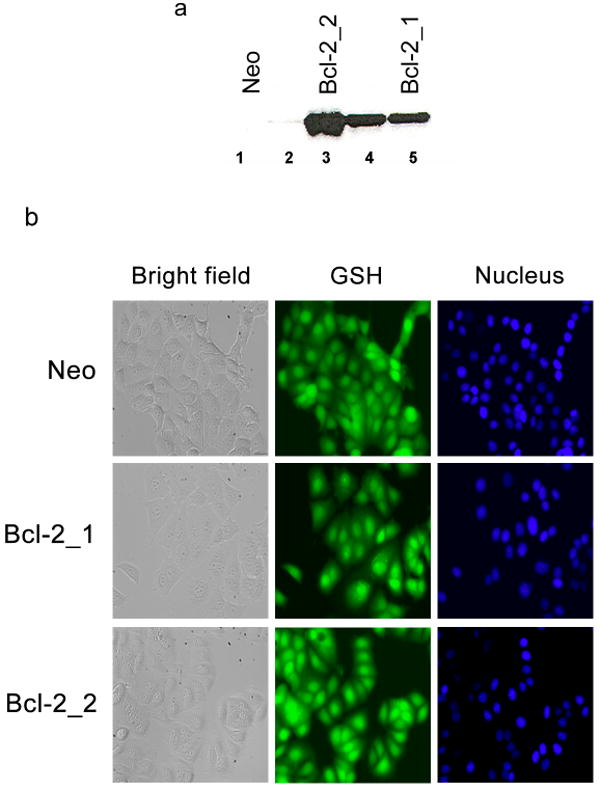
Figure 1. Characterization of MCF-7 clones.

(a) Western blot of MCF-7 clones. Lane 1 and 2, Bcl-2 expression levels in control clones transfected with empty Neo plasmid; lanes 3–5, Bcl-2 expression levels in clones transfected with a human Bcl-2 plasmid. (b) Subcellular distribution of GSH in MCF-7 clones. Cellular GSH stained with CMFDA and nuclei with Hoechst 33342. Size bar = 25 μm.
