Figure 4.
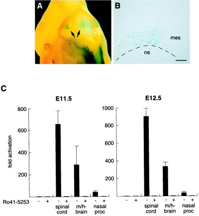
Retinoid signaling in the ventral midbrain/hindbrain region. (A) Dorsal view of a X-Gal-stained transgenic E11.5 embryo expressing UAS-hsp-gRAR and UAS-hsp-lacZ; lacZ staining in the midbrain/hindbrain region is evident (arrows). (B) Coronal section of the midbrain/hindbrain region (left side). β-Galactosidase-positive cells are located in cephalic mesenchyme, adjacent to the isthmic/pontine neuroepithelium. (Bar = 100 μm.) (C) Explant experiments of E11.5 (Left) and E12.5 (Right) wild-type embryonic tissues to detect retinoid production. JEG-3 cells transfected with CMV-gRAR and UAS-tk-luc effector and reporter plasmids were incubated overnight either alone or with indicated explants in the absence (−) or presence (+) of the RAR antagonist Ro 41-5253. Luciferase induction is shown as mean fold activation (±SD) of triplicate values. ne, neuroepithelium; mes, mesenchyme; m/h-brain, midbrain/hindbrain region; nasal proc, medial and lateral nasal processes.

