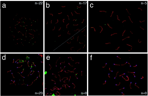
Fig. 7.
Localization of Mlh1 (a–c) and Mlh3 (d–f) on meiotic chromosomes. (a–c) Localization of Mlh1 (green foci) on pachytene chromosome spreads from WT (a) and Mlh1G67R/G67R (b and c) males reveals a dramatic decrease in frequency and intensity of Mlh1 staining in the Mlh1G67R/G67R mutant animals. Many cells show a complete absence of Mlh1G67R staining (data not shown), but others show distinct Mlh1G67R reduction as exemplified in b and c. (d–f) Mlh3 staining (green foci) of WT (d) and Mlh1G67R/G67R (e and f) males reveals a complete absence of Mlh3 staining in pachytene spreads from Mlh1G67R/G67R mutant animals. (See also larger images in SI Fig. 13 in SI Appendix). Synaptonemal complexes are stained with anti Sycp3 antiserum (red), and centromeres are detected by CREST autoimmune serum (blue).

