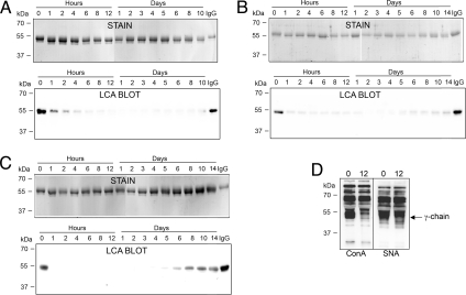
Fig. 3.
EndoS hydrolyzes IgG in vivo in rabbits. (A) SDS/PAGE (stain) and lectin blot analysis (LCA blot) of purified IgG from serum samples withdrawn from a representative rabbit at indicated time points after the first i.v. injection of 500 μg of EndoS (0 days). (B) SDS/PAGE (stain) and lectin blot analysis (LCA blot) of purified IgG from serum samples withdrawn from the rabbit at indicated time points after a second administration (35 days) of EndoS. (C) SDS/PAGE (stain) and lectin blot analysis (LCA blot) of purified IgG from serum samples withdrawn from the rabbit at indicated time points after a third administration (135 days) of EndoS. (D) Lectin blot analysis of total serum taken before (0) and 12 h (12) after the first injection of EndoS. ConA, Concavalin A; SNA, Sambucus nigra lectin. Arrow to the right indicates the position of the γ-chain of IgG.

