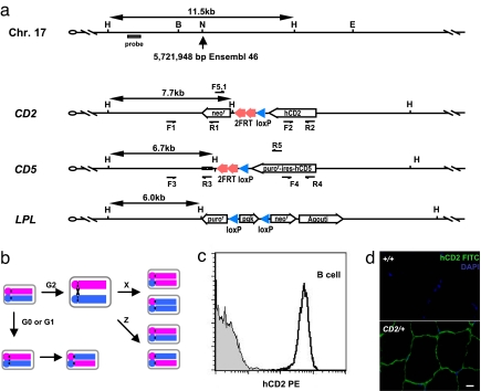
Fig. 1.
Modifications of chromosome 17 for mitotic recombination. (a) Schematic diagram of the wild-type, CD2, CD5, and LPL alleles around the insertion site of chromosome 17. The precise location of insertion site is based on Ensembl release 46. The hCD2 and hCD5-ires-puror markers are transcribed by the β-actin promoter, and the agouti marker is transcribed by the K14 promoter. F1/R1, F2/R2, F3/R3, F4/R4, and F5.1/R5 are primer pairs for evaluation of recombination across the LoxP or FRT sites. Restriction sites are as follows: B, BglII; E, EcoR V; H, HindIII; N, NheI. (b) Conceptual overview of the mitotic recombination system. Although recombination may occur in the G0, G1, and G2 phases of the cell cycle, only G2 recombination followed by X segregation will produce daughter cells carrying the segregated alleles. (c) Expression of hCD2 in lymph node B cells. Gray area is from a wild-type control mouse, and the dark line indicates data from a CD2/+ mouse. (d) Detection of hCD2 expression in the skeletal muscle of a CD2/+ mouse. (Scale bar: 10 μm.)

