Fig. 3.
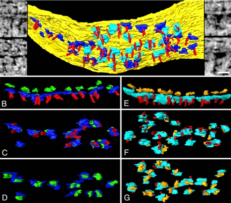
Transmembrane structures at the PSD. (A) Cytoplasmic surface of the membrane (yellow) at the PSD, showing AMPAR-type (blue) and NMDAR-type cytoplasmic domains (cyan), and vertical filaments (red). (Left Insets) AMPAR-type structures in virtual sections from the tomographic reconstruction. Arrow points to a transcleft filament. (Right Insets) NMDAR-type structures. Arrow points to a transcleft filament, and double arrow (lower right) indicates the extent of synaptic cleft. (Scale bar: 20 nm.) (B–D) AMPAR-type structures shown in cross-section (B, extracellular domain green), en face from inside the spine (C), and en face from outside the spine (D). Cytoplasmic domains of AMPAR-type structures are contacted by vertical filaments (C). (E–G) NMDAR-type structures shown in cross-section (E, extracellular domain gold), en face from inside the spine (F), and en face from outside the spine (G). Cytoplasmic domains of NMDAR-type structures are contacted by one or two vertical filaments (F).

