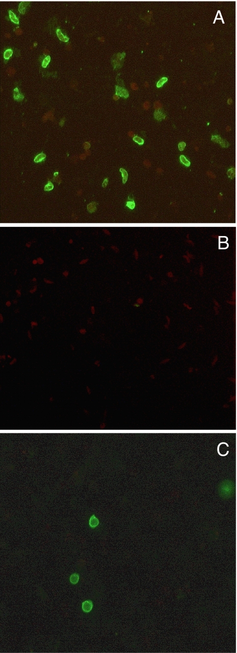
Fig. 5.
IFA and SIFA analysis of sera from mice immunized with M-Pfs10C. (A and B) Immunofluorescence microscopy with anti-M-Pfs10C mouse sera on P. falciparum gametocytes air-dried on a multispot slide (IFA) with wild-type parasites (A) and Pfs48/45 knockout parasites (B). (C) SIFA using live intact macrogametes/zygotes. (Magnification: ×400.)

