Fig. 4.
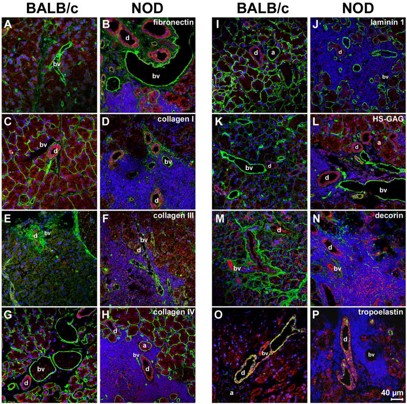
Immunohistochemical analysis of LGs of 18 weeks old NOD mice reveals massive inflammatory infiltrates, mainly located around the acini (a), blood vessels (bv) and ducts (d), associated with a spatial degradation of ECM proteins. Images show F-actin staining (red) as well as positive staining (green) for: fibronectin (A, B); collagen type I (C, D); collagen type III (E, F); collagen type IV (G, H); laminin 1 (I, J); HS-GAG (K, L); decorin (M, N); and tropoelastin (O, P). DAPI-staining was performed to show cell nuclei (blue).

