
Figure 2. Possible arrangements of the three proteolipid subunits (c, c′ and c″) in a six-membered proteolipid ring of the yeast V-ATPase.
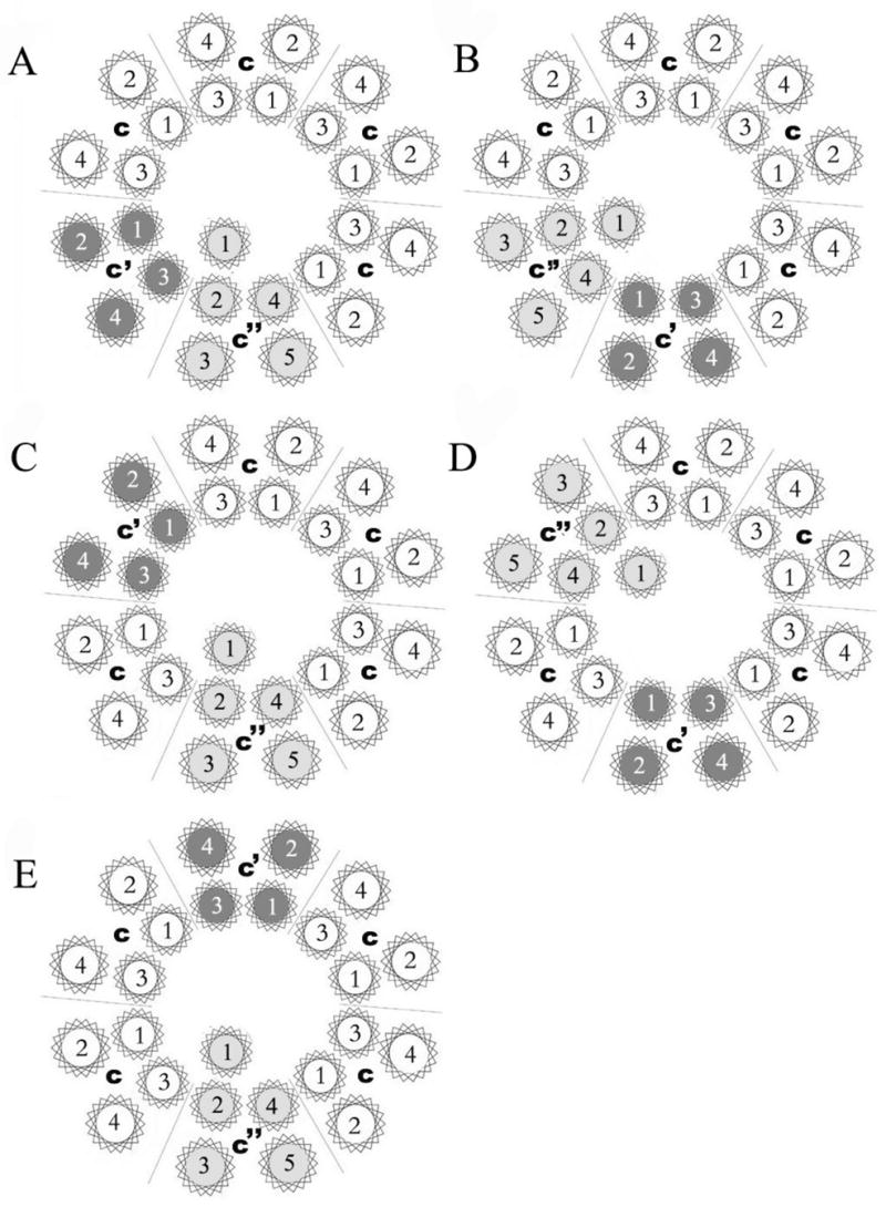
Subunit c is shown in white, subunit c′ is shown in dark gray and subunit c′′ is shown in light gray. The proteolipid ring is viewed from the lumenal side of the membrane, with the arrangement of helices within each subunit modeled after that observed in the c ring of the Na+ V-ATPase from Enterococcus hirae (27). A five TM model of subunit c″ is assumed in this diagram.