Figure 1.
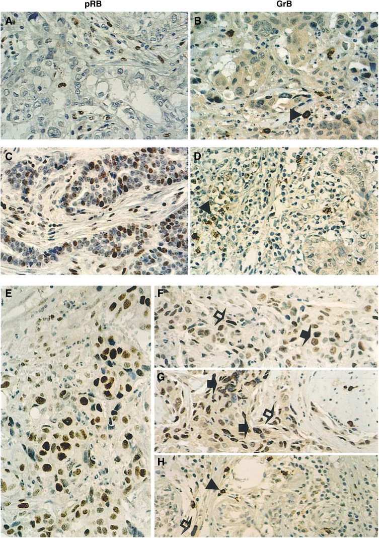
Detection of endogenous GrB in primary breast carcinomas overexpressing pRB (pRB++) by immunohistochemical staining of paraffin-embedded tissue sections. (A, C, E) pRB staining, showing typical pRB− (A), pRB+ (C), and pRB++ (E) tumours. Note that the tumour cells in panel E (pRB++) display uniformly strong pRB staining, while the tumour cells in panel (C) (pRB+) show nuclear staining heterogeneity of the RB protein, ranging from quite positive to seemingly negative (Xu, 1995). (B, D, and F–H) The same tumours corresponding to the left panels were stained for GrB. Panels B and D, in either pRB− (B) or pRB+ (D) tumours, malignant cells are GrB negative, but some infiltrating lymphocytes are GrB+. Panels (F–H), representative areas of the same pRB++ tumour shown in Panels E. GrB+ tumour cells (F, G), or lymphocytes (H) were evident. Note the finely granular distribution of endogenous GrB protein in tumour cells of panel (G). Arrowheads, GrB+ lymphocytes; solid arrows, GrB+ tumour cells; open arrows, GrB; mesenchymal and endothelial cells. Scale bar, 50 μm.

