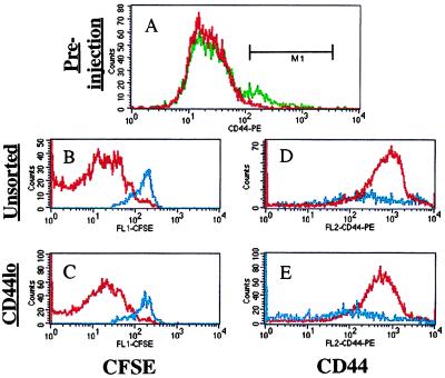
Figure 5.
Expansion of sorted CD44low “naïve” OT-I T cells is similar to that of unsorted cells. OT-I CD8+ donor cells were used as a bulk population or after sorting for low CD44 expression. Reanalysis of these populations (A) revealed the unsorted cells (green line) to be 8.7% CD44high, whereas the sorted cells (red line) were <0.7% CD44high, using the marker shown. The unsorted OT-I cells (B and D) and sorted CD44low (C and E) population were transferred into irradiated B6 (red lines) or TAP0/0 (blue lines) and assayed for CFSE expression (B and C) and CD44 staining (D and E) at day 10 after transfer. Data are representative of 2–3 mice per group.

