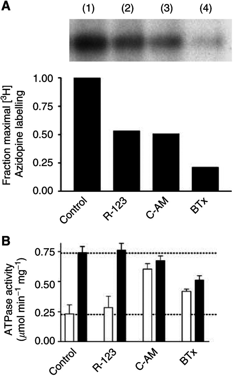
Figure 1.
Azidopine displacement by model fluorescent substrates of P-gp. (A) Purified, reconstituted human P-gp (250 ng) was incubated with [3H]azidopine (0.5 μM) for 60 min in the absence (lane 1) or presence of 10 μM rhodamine 123 (lane 2), 10 μM calcein-AM (lane 3) or 5 μM BODIPY-taxol (lane 4). The samples were then placed on ice and irradiated (315 nm, 100 W at 5 cm) for 5 min. Protein was separated from unbound [3H]azidopine by SDS–PAGE and subjected to autoradiographic analysis. (B) The ATPase activity of purified, reconstituted human P-gp (250 ng) was determined by measurement of liberated inorganic phosphate. Basal and drug-stimulated (10 μM nicardipine) ATPase activities were determined in the absence or presence of rhodamine 123 (10 μM), calcein-AM (10 μM) or BODIPY-taxol (5 μM). Basal and nicardipine-stimulated activities are represented by clear and filled bars, respectively. Error bars denote the s.e.m. and the dotted lines represent the level of basal and stimulated activity in the absence of fluorescent allocrite.

