Figure 1.
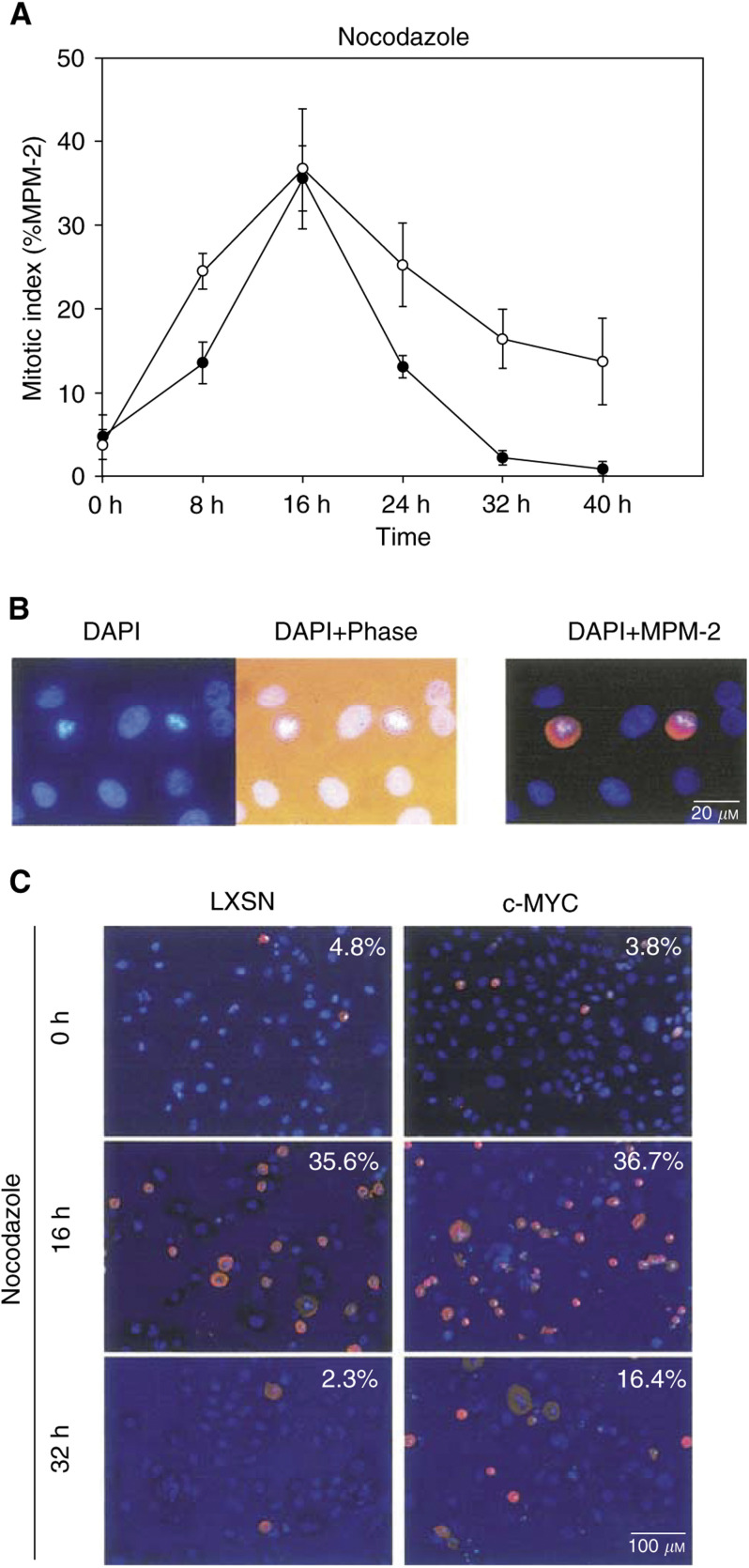
c-Myc has no effect on the time-course of nocodazole-induced mitotic arrest of normal HMECs. Following an infection with LXSN vector-only or c-Myc retroviruses, cells were allowed to express exogenous genes for 48 h. (A) Cells were treated with nocodazole, an MT destabiliser, and their degree of arrest in mitosis (%MPM-2 positive) was measured over a time-course of 0, 8, 16, 24, 32, and 40 h time-points. Both LXSN-infected controls (filled circle) and c-Myc-overexpressing cells (nonfilled circle) arrested at mitosis over the same time-course, following artificial abrogation of mitotic spindles. (B) DAPI staining identifies mitotic cells, with typical, condensed chromosomes. Rounded-up cells, on phase-contrast microscopy, match with cells having condensed chromosomes on DAPI staining. Finally, mitotic cells were confirmed with a mitosis-specific marker, MPM-2. (C) Representative figures demonstrate gradual changes of MPM-2-positive mitotic cells, following prolonged treatment with nocodazole.

