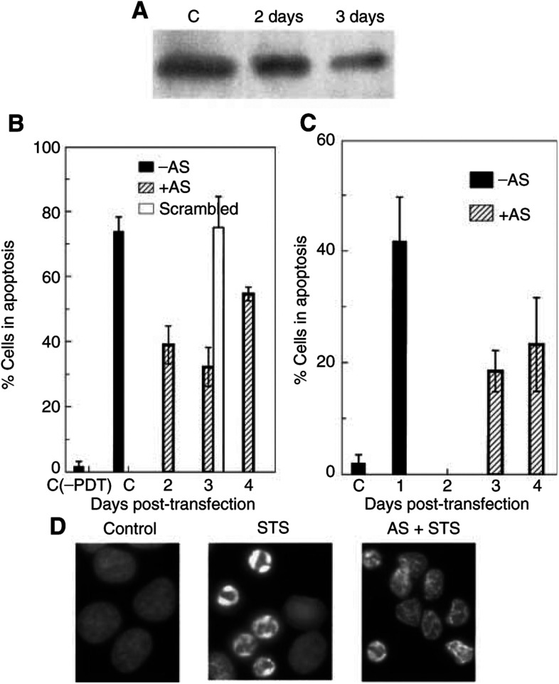
Figure 2.
Suppression of Bax expression protects MCF-7c3 cells from PDT-induced apoptosis. (A) Western blot analysis of the levels of Bax after treatment with Bax AS for 2 or 3 days. (B, C) Levels of apoptosis induced by 1 μM STS (B) or PDT (C) in MCF-7c3 cells transfected with Bax-AS or scrambled sequences. At 1–4 days after transfection, cells were exposed to PDT or STS, and 6 h later, cells were stained with Hoechst 33342 and apoptotic cells were counted. (D) Nuclear morphology of Bax AS-treated MCF-7c3 cells 6 h after STS treatment.

