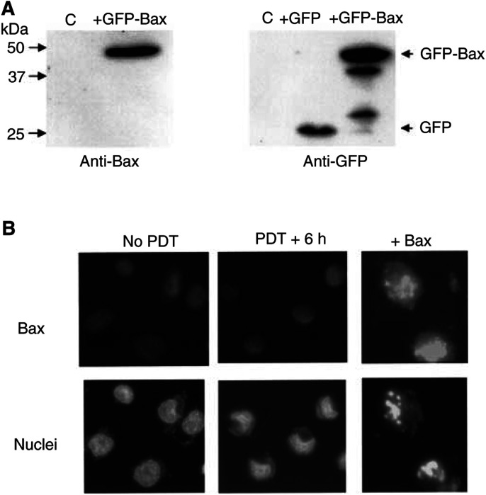
Figure 4.
Expression of Bax in DU-145 cells restores apoptosis. (A) Western blot analysis of the levels of Bax in DU-145 cells 20 h after transfection with a plasmid encoding GFP-Bax or GFP. Blots were probed with anti-Bax (left panel) or anti-GFP (right). (B) Immunocytochemical detection of Bax expression (upper panels) and nuclear morphology (lower panels) of DU-145 cells. Nontransfected cells were either untreated (left panels) or PDT-treated and examined 6 h later (middle panels). Other cells were examined 20 h after transfection with a Bax-expression plasmid.

