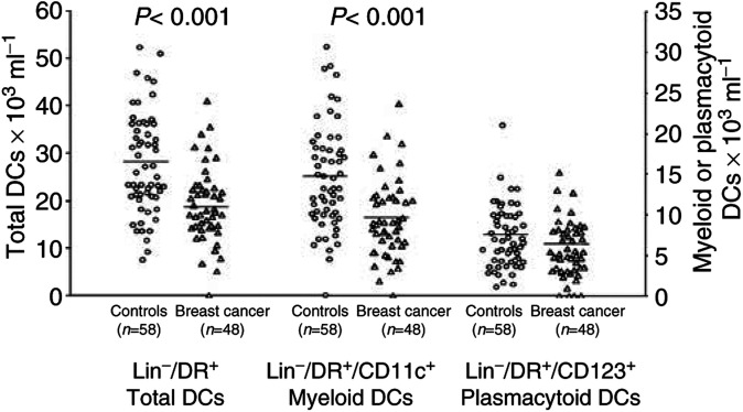
Figure 1.
Absolute number of PB DCs in breast cancer patients compared with controls. A significant decrease in PB Lin-/HLA-DR+ cells (left axis) and myeloid DCs (right axis) was observed in cancer patients. Whole blood was stained with a cocktail of FITC-conjugated mAbs recognising CD3, CD14, CD16, CD19 and CD20, and with PerCP-conjugated anti-HLA-DR mAb; myeloid DCs were identified as lineage-/HLA-DR+/CD11c+ cells, plasmacytoid DCs (right axis) as lineage-/HLA-DR+/CD123+ cells. Absolute DC counts were then determined indirectly by multiplying the percentage of DCs in the mononuclear gate times the sum of the lymphocyte and monocyte determined on a differential blood cell counter. Each symbol represents a single sample. Open circles: control subjects; open triangles: breast cancer patients. Mean values represented by horizontal lines in each series. P-values were determined using the t-test for independent samples, patients compared with controls.

