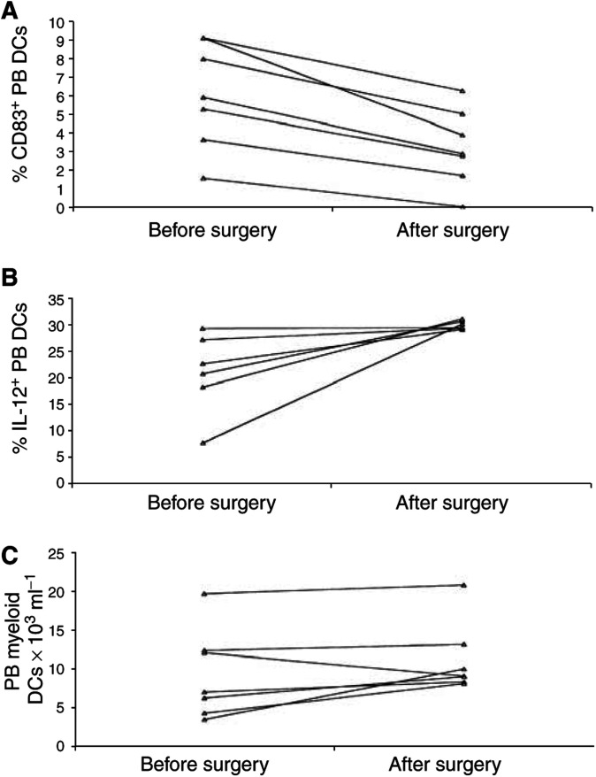
Figure 6.
Effects of surgical removal of the tumour on PB DCs. At 4 weeks after surgery, (A) the percentage of CD83+ PB DCs significantly decreased (P=0.001), with a reduction of mature PB DCs observed in all the patients; (B) the percentage of PB DCs expressing IL-12 upon LPS stimulation significantly increased (P=0.042), with a complete normalisation observed in all the patients; (C) the number of myeloid DCs in the peripheral blood of the patients only slightly increased (P=n.s.). Methods as described in Figures 2–4 Each symbol represents a single sample. P-values were determined using the t-test for paired samples.

