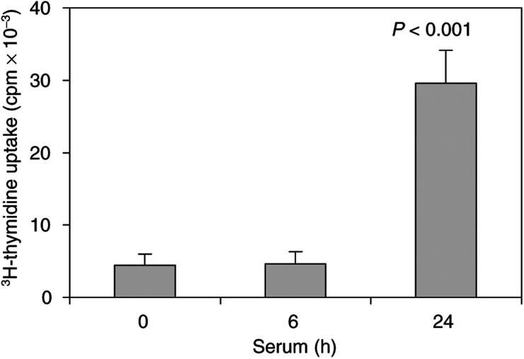
Figure 1.
3H-thymidine uptake in serum-treated hepatocytes. Cells were grown in 96-well plates in DMEM containing 10% FBS. After 24 h, cells were made quiescent by 48 h serum deprivation and then cells were either untreated (0 time point) or were treated with 15% FBS for 6 or 24 h. Cell proliferation was monitored by adding 0.5 μCi of 3H-thymidine to each well for another 3 h. The radioactivity of collected cellular DNA was determined by scintillation counting. Three independent experiments were performed and the results represent means±s.d. of radioactivity counts expressed in c.p.m.

