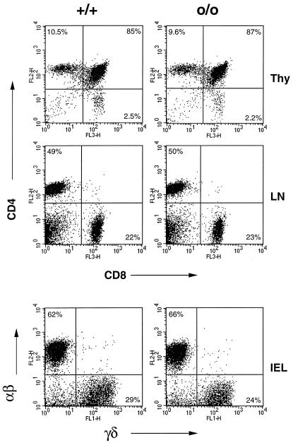
Figure 3.
HFE-deficient mice harbor normal number of various T cell subpopulations. Shown is a representative FACS analysis of lymphocytes isolated from HFEo/o and HFE+/+ littermates (13 weeks of age). CD4/CD8 staining of thymocytes and lymph node cells is shown above and αβ (TCRβ)/γδTCR staining of IELs below. Total cell numbers isolated from each mouse were similar, and the percentages of relevant populations are noted.

