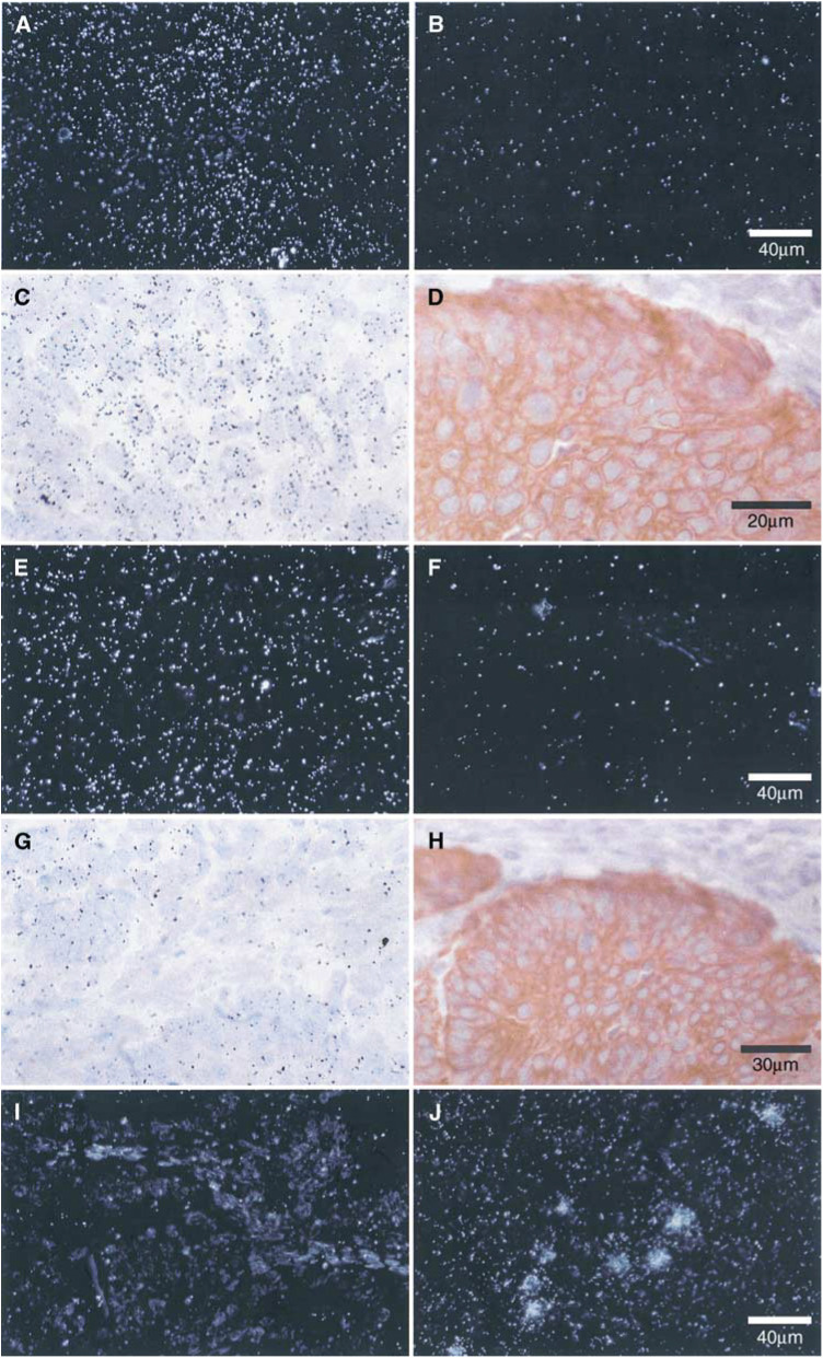
Figure 2.
Expression of mRNA encoding VEGF-related factors in the epithelial cells of endometrioid endometrial carcinoma and ACH. In situ hybridisation with VEGF-A antisense probe in a moderately differentiated (FIGO grade 2) carcinoma shown under dark-field (A) and light-field (C) conditions shows hybridisation in epithelial carcinoma cells. There is no specific hybridisation when a sense probe (negative control) is used (B). The scale bar in (B) applies to (A), (B), (E), (F), (I) and (J). Immunostaining of a serial section with anti-human cytokeratin confirms the epithelial localisation of silver grains (D) (scale bar applies to ACH (C) and (D). In situ hybridisation with VEGF-B antisense probe in ACH shown under dark-field (E) and light-field (G) conditions shows diffuse hybridisation in epithelial and stromal cells. There is no specific hybridisation when a sense probe (negative control) is used (F). Immunostaining with anti-human cytokeratin (H) identifies the epithelial and stromal cells (H) (scale bar applies to (G) and (H). In situ hybridisation with VEGF-C antisense probe in a poorly differentiated (FIGO grade 3) carcinoma shown under dark-field conditions (I) shows no specific hybridisation when compared with a human B-cell lymphoma (J).

