Figure 8.
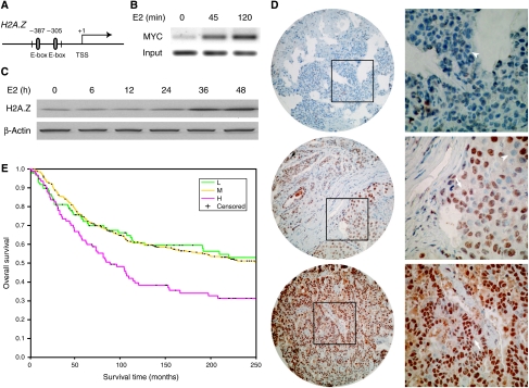
H2A.Z is a target of the E2 cascade and is associated with breast cancer progression. (A) The H2A.Z promoter harbors two canonical E-boxes (5′-CACGTG-3′), which serve as MYC response elements. (B) ChIP-PCR demonstrates that MYC is recruited to the H2A.Z promoter in MCF7 cells after E2 (10 nM) exposure. (C) Western blot demonstrates the induction of H2A.Z protein level in MCF7 cells in response to E2 (10 nM). (D) Immunohistochemical staining of tissue arrays for H2A.Z protein. Tissue microarrays were stained with an antibody specific for H2A.Z (brown and arrows) and counterstained with hematoxylin (blue and arrowheads). Three representative tumor samples with differing H2A.Z protein levels are shown (from upper to lower panel: low, moderate, and high staining). For each tissue shown on the left (× 60 magnification), the inset is highlighted in the corresponding right panel (× 180 magnification). (E) Kaplan–Meier curves of overall patient survival for 517 breast cancer patients with corresponding variations in H2A.Z tumor expression (low (L): 98 patients; moderate (M): 326 patients; and high (H): 93 patients). The P-value is calculated based on the log-rank test.

